Scientists invent new 3D imaging analysis technology

According to a report published in the Science Report on the 17th, according to a report published in the Science Report on the 17th, the engineers, doctors and radiologists from the University of Cambridge in the UK have invented a new The use of 3D imaging analysis techniques to detect small changes in arthritis will help to better understand the pathogenesis and process of osteoarthritis and develop more accurate treatments without the need for invasive tissue sampling.
Dr. Tom Turmezei of the Department of Engineering at the University of Cambridge said: "This technology is a valuable tool for analyzing arthritis in both clinical and research, and it can be used in conjunction with 3D statistical analysis to accelerate the development of new therapies."
At present, there is no recognized method for treating osteoarthritis, which is a common arthritis caused by the wear of articular cartilage at the end of the bone. Artificial joint replacement is the only treatment.
Turmezei said, “Using this technology, we hope to identify osteoarthritis as early as possible and find potentially effective treatments before it worsens. It can be used to screen high-risk groups, such as those known to have arthritis or have previous joints. human damage, but also those elite athletes due to the joint under constant pressure and arthritic. "
The most commonly used detection methods at present are radiation, magnetic resonance imaging and ultrasound. Doctors can also use two-dimensional X-rays to diagnose osteoarthritis by identifying areas where articular cartilage wears down. However, this imaging technique is not precise enough to detect subtle changes in joints.
Turmezei pointed out that in addition to the lack of accuracy, 2D X-ray results still rely on manual analysis and interpretation. The latest technology will make up for these shortcomings, and it has the ability to observe subtle changes in joints, and has never been able to diagnose early diseases more accurately.
This new technique, known as the "joint space mapping" method, uses images from standard computed tomography (also known as CT scans), which are typically not used for joint monitoring. This technique primarily analyzes CT images to identify subtle changes in the inter-bone space of the joint.
After testing, the researchers found that the imaging accuracy of this new technology is twice that of current X-ray combined imaging. But the researchers also said that although CT scans have not yet been approved for monitoring joints, the combination of low-dose CT scans and 3D imaging techniques will hopefully lead to more effective treatment decisions or new treatment options for patients, as well as bone and joints. Inflammatory care has brought better development prospects.

Flower Type LED Operating Lamp

The flower type surgical lamp is designed at 2016, when it was been pushed into the market, it has good echo from the customers, because the flower type surgical lamp outlook is different with normal round types lamps, its outlook is just like a flower, it is very beautiful , also can add beauty in the operation room, the flower type ot lamp change the normal condition for baldness operation room style, can make doctor and patients feel much difference; for the flower type surgery lamps specification, it is almost same as round type surgical lamp, in the first year, it also has good selling;

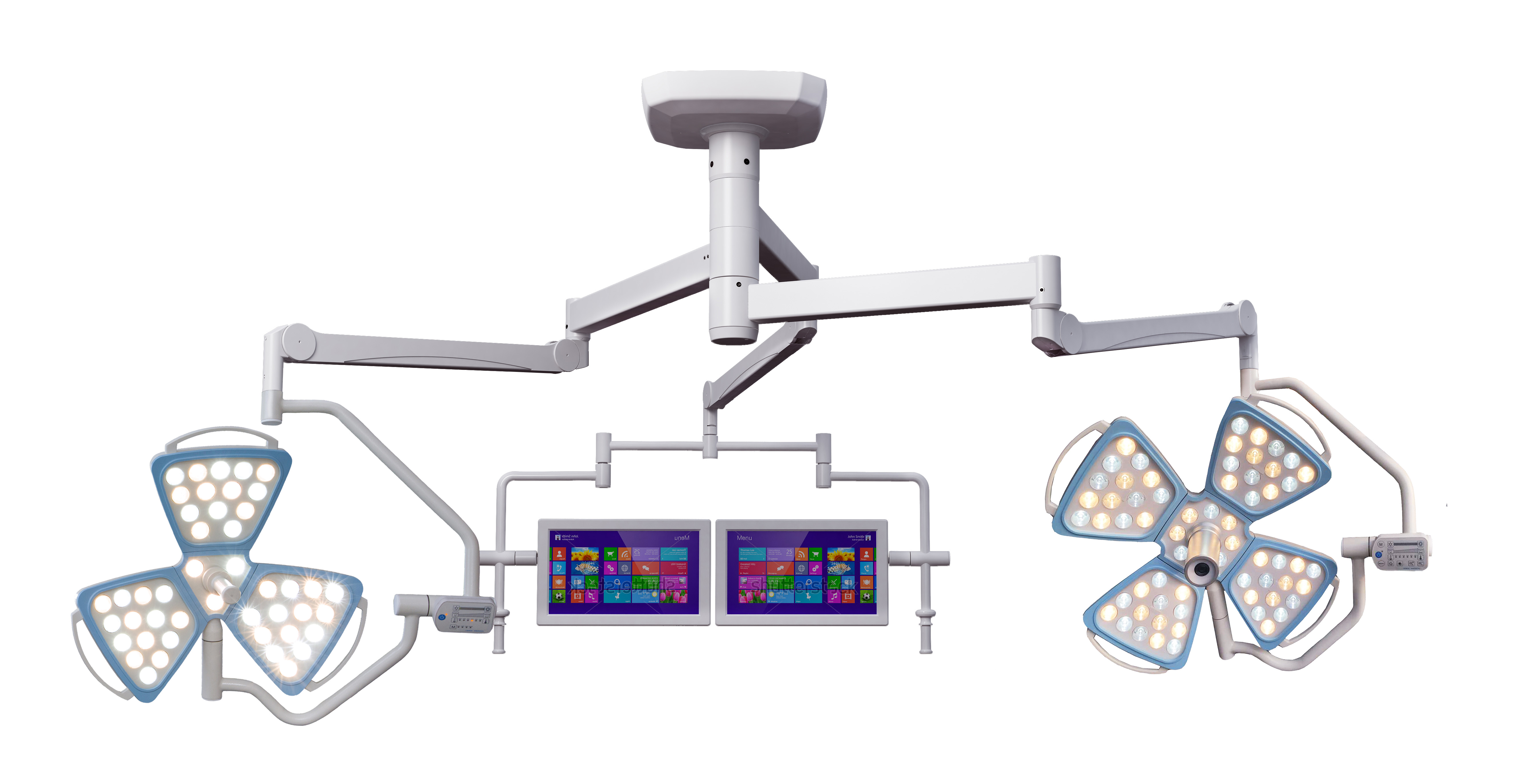
Flower type surgical lamp also has three types, ceiling types which include single dome, double dome , with camera or without camera; wall type and mobile type flower operation lamp;


3300


LED Ot Lamp,LED Operation Lamp,LED Surgery Light,LED Surgical Light

Shandong Lewin Medical Equipment Co., Ltd. , https://www.lewinmed.com