The past and present of the blood cell counting board
INTRODUCTION <br> The blood cell counting board has been a must-have tool for doctors and biologists for more than 100 years. It was originally used by doctors to study blood samples from patients and thus created the field of research in hematology. In the eighteenth and early nineteenth centuries, the blood cell counting board experienced a series of significant developments. Jack David Davis gave a detailed introduction to the development of the blood cell counting plate in the paper "The Hemocytometer and Its Impact on Progressive-Era Medicine" [1]. Here we provide you with a brief introduction to Davis's thesis, so that readers can understand the past and present of the blood cell counting board.
Development of blood cell counting board
In 1852, German scientist Karl Vierdordt (1818-1884) invented the first method to accurately count red blood cells. He uses a capillary with an inner diameter and length of 0.1-0.2 mm and 5-8 mm to draw a blood sample; after the sample is loaded into the capillary, the capillary diameter and sample length can be measured to obtain an accurate sample volume; then the blood sample is applied to The egg whites were coated on a glass plate and allowed to air; finally, the cells were counted using a microscope equipped with a graduated eyepiece. Although this method is time consuming and laborious, very accurate counting results can be obtained.
Twenty-two years later, in 1874, Louis Charles Malassez (1842-1909) invented a new method of cell counting. As shown in Figure 1, he bonded the flat capillary to the glass plate as a counting chamber. A scale on the glass plate converts the capillary length to the sample volume. Cell concentration can be obtained by counting the number of red blood cells in the capillary and multiplying by the dilution factor [1, 2].
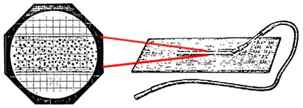
Figure 1. Capillary cell counter designed by Louis Charles Malassez
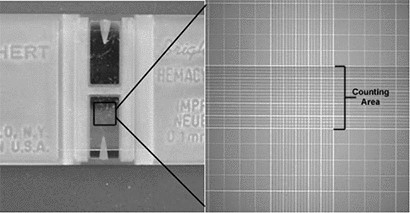
In 1875, Georges Hayem (1841-1933) invented another counting board. He glued a piece of 0.2 mm thick glass with a 1 mm diameter hole on the glass plate (pictured). A drop of blood is added directly to the well and the coverslip is covered. Cell counts were then performed using a microscope with a scale (0.2 mm x 0.2 mm) and the cell concentration was calculated by combining the hole height (0.2 mm) (Figure 2). Since the sample is loaded without siphoning, the distribution of cells is not uniform. Although Hayem manufactures and sells his products, it is not widely accepted because of the cumbersome preparation steps and the problem of poor count consistency [1, 2].
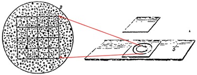
Figure 2. Cell counting board designed and manufactured by George Hayem
In 1877, William Gowers (1845-1915) improved Hayem's counting board, simplifying the counting method. Because the previous counting method required a special microscope (with an eyepiece that calibrated the scale), it was very inconvenient to use. William and Hawksley teamed up to invent the first counting chamber with a ruler directly on the glass. The ruler is designed as a 0.1 mm x 0.1 mm square with a measuring chamber thickness of 0.2 mm, which allows accurate determination of sample volume and cell concentration (Figure 3). In 1906, William optimized the scale to a long square of 0.1 mm x 0.2 mm. But because it was not published in industry journals, William's counting board was not widely accepted [1, 2].
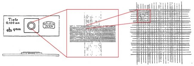
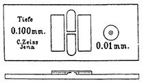
The next improvement was completed by Richard Thoma (1847-1923) in 1881, mainly for the setting of the height of the counting chamber and the increase of the grid. He first glued a piece of glass with a 11 mm diameter hole in the middle to the glass plate, and then glued a 5 mm diameter glass disc to the center of the hole. The outer glass sheet is 0.1 mm thicker than the inner glass sheet, so a counting chamber with a height of 0.1 mm is naturally formed. The area of ​​1 mm 2 at the center of the bottom of the glass plate is divided into 16 squares, each of which is subdivided into 25 small squares; each square has a size of 0.25 mm×0.25 mm (Fig. 4). This counting plate is manufactured by Carl Zeiss and Jena.
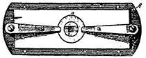
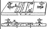
In 1884, Russian scientist Sergei Alferow designed a new type of counting plate. Alferow's counter plate separates the counting chamber from the plate by two parallel grooves. The height of the counting chamber is controlled by four scaled screws, and the sample is added to the counting chamber by siphoning to increase the uniformity of cell distribution in the counting chamber (Figure 5). Alferow was the first person to microscopically photograph cells in a counting plate. He projects the photo on a glass plate with scales and then identifies and counts each cell with a pencil. He believes his method can get more accurate results, because the photomicrograph is the most realistic record of the cell status in the counting plate [1, 2].
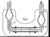
In 1903, W. Brünings invented a counting plate that integrated the sample and the sample. He connected the counter plate to a mixing chamber with a pipette. When the blood sample is mixed in the pipette, it is transferred directly to the counting chamber (Figure 6). This kind of counting board can be called the earliest application of "chip lab technology" [1, 2].
The biggest contribution to improving the ease of use and precision of the blood cell counting plate is Karl Bürker (1872-1957). Bürker's counting plate consists of a heavy glass plate and three platforms glued to the glass plate. The three platforms are arranged in parallel on the glass plate; the middle platform (25 mm x 5 mm) is divided into two parts by a 1.5 mm wide groove with rounded ends; the platforms on both sides (21 mm x 7.5 mm) are in the middle The platform is 0.1 mm high and is separated from it by a 1.5 mm wide channel. After the coverslips are placed on the platforms on both sides, a depth of 0.1 mm is formed. The two parts of the intermediate platform each have a ruler with a scale of 1 mm 2 and are divided into 400 small squares. Bürker's counter plate loads the sample by capillary action; there are two counting chambers on the counter plate, and the sample can be counted repeatedly without cleaning (Figure 7). Through this series of successful improvements, Bürker's counting board became the most commonly used type of blood cell counting board before 1921 [1, 2].
Improvements in the hemocytometer grid <br> The grid of blood cell countboards has undergone several improvements. Thoma's 1879 design grid laid the foundation for early blood count panels. But with the development of hematology research, scientists have found that such a grid is not suitable for white blood cell detection because white blood cells are larger than red blood cells and platelets.
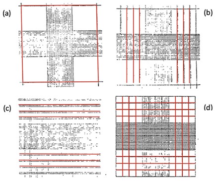
In 1892, J. Zappert expanded the mesh area to 9 mm 2 and consisted of nine 1 mm 2 areas. In 1894, A. Elzholz added three sets of vertical lines to the left and right sides. In 1902, W. Türk added three more horizontal lines to Elzholz. The grid at this point is similar to today's blood cell counting boards. In 1907, O. Neubauer changed the double line to a single line, further simplifying the design of the grid (Fig. 8) [1].
Summary <br> After a century of improvement, the blood cell counting board has evolved from its embryonic form to a precision tool that is common worldwide today (Figure 9). Although there are different models for blood cell counting plates, it is still the gold standard for cell counting in both experimental and clinical diagnostic fields. In the next article, we will share with you the source of error in cell counting and the method of reducing errors using a hemocytometer to improve cell count and accuracy of downstream experiments.
references
1. Davis, JD, THE HEMOCYTOMETER AND ITS IMPACT ON PROGRESSIVE-ERA MEDICINE, in Department of History 1995, University of Illinois at Urbana-Champaign: Urbana. p. 268.
2. Verso, ML, Some Nineteenth-Century Pioneers of Haematology. Medical History, 1971. 15(1): p. 55-67.
3. Rouge, M. Counting Cells with a Hemacytometer. 2002; Available from: http://
Nexcelom and Daktronics are
Founded in 2003 in Boston, USA, Nexcelom Bioscience is a leader in the development and commercialization of high performance cell quantitative analysis platforms. As a global company. Since the successful development of the first cell counter, the Cellometer Auto T4 in 2005, and the recognition of NIH customers, the Cellometer brightfield, fluorescence and image flow cell quantitative analyzer series has been developed.
In 2014, it successfully acquired the Celigo full-field in-situ cell analyzer product line from Brooks Life Sciences, quickly integrating and enhancing the company's R&D capabilities and customer application support capabilities in cell quantification technology.
Cellometer and Celigo are widely used in basic life science research such as new drug development, immunotherapy, vaccine development, drug evaluation, and process development such as brewing and bioenergy. At present, the number of installed machines in the world has reached more than 5,000, and more than 3,200 articles have been cited in peer-reviewed literature.
Dakota is a biological as Nexcelom company's distributor in China, and Nexcelom joined forces with a strong technical team in China to provide professional demo presentations, as well as after-sales support and maintenance.
Fried Dough Twist,Crispy Dough,Fried Snack Food,Fried Hemp Leaves
Zhejiang Shanying Trading Co.,Ltd. , https://www.shanyingtrading.com